Empyema thoracis is an accumulation of pus in pleural space. It is most often associated with pneumonia due to Streptococcus pneumoniae, although Staphylococcus aureus is most common in developing nations and Asia [1]. Haemophilus influenzae, group A Streptococcus, gram negative organisms, tuberculosis, fungi, malignancy and trauma are other causes. Empyema thoracis consists of three stages-
1. Exudative phase (fibrinous exudates forms on pleural surfaces),
2 Fibrinopurulent phase (fibrinous septa form, causing lobulation and thickening of parietal pleura), and
3. Organization phase.
Empyema thoracis is an uncommon complication of childhood pneumonia and general pediatricians may only see a few cases in their career [2]. Although mortality rates in pediatric empyema thoracis are very low, empyema thoracis causes significant morbidity including substantial health care costs and burden of care. Childhood empyema thoracis occurs in 0.7 – 3.3 per 100,000 population worldwide. If pus is not drained in second phase it may dissect through pleura into lung parenchyma leading to bronchopleural fistula (BPF), pyopneumothorax, in abdominal cavity or through chest wall (empyema necessitans). If organized, lung may collapse and become surrounded by thick inelastic peel.
Computed tomographic scan with contrast enhancement should be performed with lung and mediastinal windows to reveal the exact extent and nature of the disease. Very few authors have realized the importance of CECT chest while deciding for surgery.[3] The majority of studies that we reviewed were based on a chest radiograph and not a CT scan leading to incorrect judgment of the stage of the disease as well as delay in surgical intervention with consequent increased morbidity[4]. A chest radiograph provides only 2-dimensional information. It may only show opacity occupying a certain area of the hemithorax, which may be secondary to consolidated parenchyma, pleural peel or a lung abscess. On the other hand, the ability of CECT to show the thorax in various sections and planes helps to reveal precise information about the location, density and volume of the fluid along with the thickness of the pleural peel and the status of the underlying lung with its degree of entrapment.
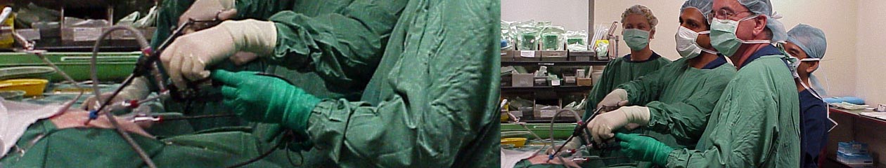
The mainstay of treatment of pyogenic pleural empyema is control of on-going infection and the prevention of recurrent infection and late restriction. The complicated pleural effusion or empyema conditions were traditionally managed through early drainage, either by means of a chest tube or through a thoracotomy and open drainage [5]. Delaying surgical treatment in these situations or, incomplete drainage of the pleural space with persistent signs of infection is responsible for functional impairment and is associated with substantial morbidity and mortality [6]. The physicians and pulmonologists prefer conventional therapies, such as antibiotics and insertion of the pig-tail catheter over surgery. This may cause late referral and further complications of the empyema cases. It is important to identify risk factors associated with patient who fail fibrinolytic therapy and who may benefit from primary mechanical debridement [7]. Operations for empyema thoracis were conventionally performed by open thoracotomy, whereas the video-assisted thoracic surgery (VATS) approach in the hands of an experienced surgeon has all the advantages of minimally invasive surgery and has similar final outcomes as open surgery. Various postoperative characteristics determine the success of VATS decortication , that is, shortening of operation time, chest tubes duration, postoperative hospital stay, and less perioperative mortality [8].
The success of video-assisted thoracoscopic surgery (VATS) may be dependent on the stage of disease, early decortication prevents permanent changes in the underlying lung parenchyma. VATS allows equally effective decortication for empyema as thoracotomy. However, the VATS approach gives less pain and greater patient acceptance.
Primary VATS is much more advantageous in children as the thoracic cavity is cleaned and loculi broken primarily before inserting an intercostal drain and allowing the infection to become chronic. It leads to better lung expansion and early recovery.
References:
- Glenna B, Lossef SV. Purulent pleurisy or empyema. In: Kliegman RM, Stanton BF, Schor NF, St Geme III JW, Behrman RE, editors. Nelson Textbook of Pediatrics. 19 ed. Philadelphia: Saunders; 2011. p. 1507.
- Strachan R, Jaffe A, Australian Research Network in E. Assessment of the burden of paediatric empyema in Australia. J Paediatr Child Health. 2009;45(7-8):431–6.
- Gun F, Salman T, Abbasoglu L, Salman N, Celik A. Early decortication in childhood empyema thoracis. Acta Chir Belg. 2007;107:225–7.
- Chan W, Keyser-Gauvin E, Davis GM, Nguyen LT, Laberge JM. Empyema thoracis in children: A 26-year review of the Montreal Children’s Hospital experience. J Pediatr Surg. 1997;32:870–2.
- Kim BY, Oh BS, Jang WC, Min YI, Park YK, Park JC. Video-assisted thoracoscopic decortication for management of postpneumonic pleural empyema. Am J Surg. 2004;188:321–4.
- Anstadt MP, Guill CK, Ferguson ER, Gordon HS, Soltero ER, Beall AC, Jr, et al. Surgical versus nonsurgical treatment of empyema thoracis: An outcomes analysis. Am J Med Sci. 2003;326:9–14.
- Espinosa CM, Fallat ME, Woods CR, Weakley KE, Marshall GS. An approach to the management of pleural empyema with early video assisted throcascopic surgery and early transition to oral antibiotic therapy. Am Surg 2016; 82: 295-301.
- Tong BC, Hanna J, Toloza EM, Onaitis MW, D’Amico TA, Harpole DH, et al. Outcomes of video-assisted thoracoscopic decortication. Ann Thorac Surg. 2010;89:220–5.
