Anorectal malformations (ARMs) are among the more frequent congenital anomalies encountered in paediatric surgery, with an estimated incidence ranging between 1 in 2000 and 1 in 5000 live births. Antenatal diagnosis of an isolated ARM is rare. Most cases are diagnosed in the early neonatal period. There is a wide spectrum of presentation ranging from low anomalies with perineal fistula having simple management to high anomalies with complex management.
Advances in the imaging techniques with improvement in knowledge of the embryology, anatomy and physiology of ARM cases have refined diagnosis and initial management. There has been marked improvement in survival of such patient over the last century.
Imaging: Cross Table Lateral pelvic radiography at 24 hours: It is better to wait 24 hours after birth to observe for possible maximal pelvic pouch distension and then to use cross-table lateral pelvic radiography with a radio-opaque marker on the anal dimple with the child in the prone position and the hips slightly raised. If the pouch is observed within 1 cm of the marker, some surgeons offer primary repair without colostomy. For pouches farther than 1 cm, colostomy is performed.
X ray abdomen: Two views of the abdomen with sacrum, posteroanterior and lateral, should be obtained to measure sacral ratios and to look for sacral defects, hemivertebrae, and presacral masses. This should be performed before surgery.
Abdominal ultrasonography: This study is specifically used to examine the genitourinary tract and to look for any other masses. Hydronephrosis, hydrocolpos, presacral mass, abdominal mass, or any similar finding can profoundly affect management.
Augmented-pressure distal colostography: This is the single most important diagnostic test used to clarify the anatomy in all children with malformations who require colostomy. The catheter is pulled back, and water-soluble contrast is injected by hand. This pressure is required to overcome the pressure of the levator muscles and to allow the contrast to flow into the lowest part of the colon and reveal any fistula. In patients with a fistula to the urinary tract, the bladder often fills, and the study is continued to obtain as much information as is provided with voiding cystourethrography. If no fistula is present, the distal pouch has a rounded appearance, and no urinary extravasation is visible.
If a urinary fistula is suspected, broad-spectrum antibiotics can be administered, although anaerobic coverage is unnecessary within the first 48 hours of life. Any cardiac murmurs identified upon physical examination should be evaluated using echocardiography prior to surgical intervention. The remainder of treatment includes diagnostics and surgical evaluation and management
The management of ARM has moved forward from classical procedures to PSARP to minimal invasive procedures. But still the fecal and urinary incontinence can occur even with an excellent anatomic repair, mainly due to associated problems.
Surgery for ARM: Usually has 3 stages until primary pull through has been performed.
Colostomy: A colostomy is performed in children who are not amenable to primary pull-through either because of malformation complexity (any urinary fistula in boys, vestibular fistula and cloaca in girls, no fistula in either sex >1 cm from perineal skin) or associated comorbidity.
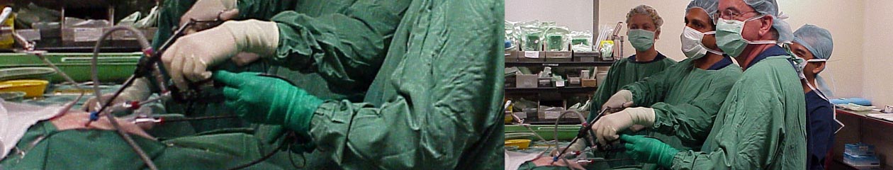
Definitive pull through: For Intermediate anomalies PSARP is the procedure of choice. For high ARM, laparoscopic assisted pull through procedure is preferred to combined abdomino-perineal pull through. Pull-through for imperforate anus offers many advantages, including excellent visualization of the rectal fistula and surrounding structures, accurate placement of the bowel through the anatomic midline and levator sling, and minimally invasive abdominal and perineal wounds.
Colostomy closure: Once the wound has completely healed and postoperative dilations have achieved their goal (ie, the neoanus is at the desired size), the colostomy may be closed in traditional surgical fashion.
