A pancreatic pseudocyst is a circumscribed collection of fluid rich in pancreatic enzymes, blood, and necrotic tissue, located in the lesser sac. Pancreatic pseudocysts are commonly complications of pancreatitis, however in children they frequently occur following abdominal trauma. Pancreatic pseudocysts account for approximately 75% of all pancreatic masses.
Symptomatic pancreatic pseudocysts are an indication that a cystogastrostomy needs to be performed. Pancreatic pseudocysts are collections of pancreatic fluid encased by a wall of non-epithelialized granulation tissue and fibrosis. They can be caused by leakage of the pancreatic duct, or as a result of inflammatory pancreatitis. Symptoms of this include abdominal bloating, difficulty eating and digesting food, and constant pain or deep ache in the abdomen. A lump can be felt in the middle or left upper abdomen if a pseudocyst is present. To further diagnose a pancreatic pseudocyst an abdominal CT scan, MRI or ultrasound can be used. Emergency surgery may need to be performed if there is a rupture of the pseudocyst. This can be detected from symptoms of bleeding, shock, fainting, fever and chills, rapid heartbeat, or severe abdominal pain.
Cystogastrostomy is a surgery to create an opening between a pancreatic pseudocyst and the stomach when the cyst is in a suitable position to be drained into the stomach. This conserves pancreatic juices that would otherwise be lost. This surgery in children is performed by a Pediatric surgeon to avoid a life-threatening rupture of the pancreatic pseudocyst.
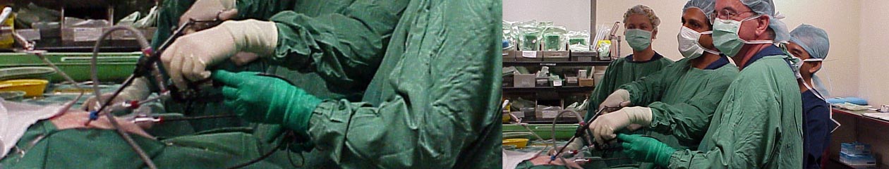
As has been a trend, more surgeons now are adopting to laparoscopic cystogastrostomy for its advantages over open surgery. After 3 ports are introduced in the umbilicus and L & R para rectal, the anterior was of stomach is opened with a harmonic shear over the most prominent site of swelling. A needle cautery is introduced and aspirated to confirm the presence of pseudocyst. An opening is made into the posterior wall of stomach withe the pseudocyst. A Endo GI stapler is fired into the posterior wall swelling which divides the posterior wall and pseudocyst together and staples the cut edges to prevent any bleed. The anterior layer of stomach is stitched back with running 3-0 vicryl to close the anterior wall opening. Port sites are closed leaving a drain in situ. Below is a video demonstration of the procedure.
