A neurofibroma is a type of nerve tumor that forms soft bumps under or on the skin. A neurofibroma may develop within a major or minor nerve anywhere in the body. This common type of benign nerve tumor tends to form more centrally within the nerve.


Symptoms are mostly mild or absent. If the tumor presses against nerves or grows within them, the patient may experience pain or numbness in the affected area. A neurofibroma is usually benign (non cancerous). Rarely, it can become cancerous (malignant).
A neurofibroma can arise with no known cause(idiopathic), or it may appear in people with a genetic condition called neurofibromatosis type 1. Diagnosis of a neurofibroma is based on a physical examination, or the results of an imaging test such as a CT or MRI scan. These imaging studies can help pinpoint where the tumor is, find very small tumors, and identify what tissues are affected or nearby. A biopsy maybe done by a radiologist before surgery to diagnose the mass as being or malignant neurofibroma.

Neurofibroma treatment usually involves monitoring or surgery.Monitoring: Observation is recommend of a tumor if it’s in a place that makes removal difficult or if it’s small and causes no problems. Observation includes regular checkups and imaging tests to see if your tumor is growing.
Surgery to remove the tumor. Symptoms can be relieved by removing all or part of a neurofibroma that’s pressing on nearby tissue or damaging organs. What type of operation is performed depends on the location and size of your tumor and whether it’s intertwined with more than one nerve. The goal of surgery is to remove as much of the tumor as possible without causing further nerve damage.
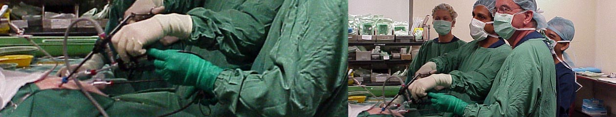
Thoracic Neurofibroma is a very rare occurrence. Below is a video demonstration of Thoracoscopic excision of neurofibroma at the thoracic inlet.
