Infantile Hypertrophic pyloric stenosis (IHPS) causes a functional gastric outlet obstruction as a result of hypertrophy and hyperplasia of the muscular layers of the pylorus. In infants, IHPS is the most common cause of gastric outlet obstruction and the most common surgical cause of vomiting.
Infants with IHPS typical present with nonbilious vomiting at 4-8 weeks of age
- Although vomiting may initially be infrequent, over several days it becomes more predictable, occurring at nearly every feeding
- Vomiting intensity also increases until pathognomonic projectile vomiting ensues
- The baby in the early stage of the disease remains hungry and sucks vigorously after episodes of vomiting
- Prolonged delay in diagnosis can lead to dehydration, poor weight gain, malnutrition, metabolic alterations, and lethargy
Careful physical examination provides a definitive diagnosis for most infants with IHPS. The diagnosis is easily made if the presenting clinical features are typical, with projectile vomiting, visible peristalsis, and a palpable pyloric tumor. Early in the course of the disease, however, some of the classic signs may be absent.
An enlarged pylorus, classically described as an “olive,” can be palpated in the right upper quadrant or epigastrium of the abdomen in 60-80% of infants [1,2].
Diagnosis
Serum electrolytes should be measured to document adequacy of fluid resuscitation and correction of electrolyte imbalances before surgical repair. The classic biochemical abnormality in HPS is hypochloremic, hypokalemic metabolic alkalosis.
Ultrasonography
- The criterion standard imaging technique for diagnosing HPS
- Muscle wall thickness 3 mm or greater and pyloric channel length 14 mm or greater are considered abnormal in infants younger than 30 days
Barium upper GI study
- Effective when ultrasonography is not diagnostic
- Should demonstrate an elongated pylorus with antral indentation from the hypertrophied muscle
- May show the “double track” sign when thin tracks of barium are compressed between thickened pyloric mucosa or the “shoulder” sign when barium collects in the dilated prepyloric antrum
- After upper GI barium study, irrigating and removing any residual barium from the stomach is advisable to avoid aspiration
Management
Hypertrophic pyloric stenosis is the most common condition requiring surgery in infancy. Correction of an associated fluid and electrolytes disturbances is vital prior to general anesthesia induction [3]. Surgical repair of HPS is fairly straightforward and without many complications. However, properly preparing the infant is vitally important.
Preoperative management
- Directed at correcting the fluid deficiency and electrolyte imbalance
- Base fluid resuscitation on the infant’s degree of dehydration
- Most infants can have their fluid status corrected within 24 hours; however, severely dehydrated children sometimes require several days for correction
- If necessary, administer an initial fluid bolus of 10 mL/kg with lactated Ringer solution or 0.45 isotonic sodium chloride solution
- Continue IV therapy at an initial rate of 1.25-2 times the normal maintenance rate until adequate fluid status is achieved
- Adequate amounts of both chloride and potassium are necessary to correct metabolic alkalosis
- Unless renal insufficiency is a concern, initially add 2-4 mEq of KCl per 100 mL of IV fluid
- Urine output and serial electrolyte determinations are performed during resuscitation
- Correction of serum chloride level to 90 mEq/L or greater is usually adequate to proceed with surgical intervention
- Before induction of anesthesia, aspirate the infant’s stomach with a large-caliber suction tube to remove any residual gastric fluid or barium; saline irrigation is occasionally necessary to remove a large quantity of barium
Surgical treatment
- Ramstedt pyloromyotomy remains the standard procedure of choice
- The usual approach is via a right upper quadrant transverse incision that splits the rectus muscle and fascia
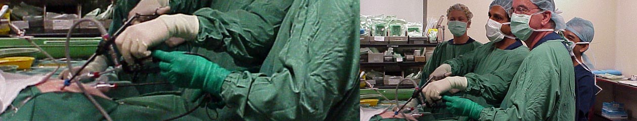
- Laparoscopic pyloromyotomy may also be used[4]. It is a viable alternative to open pyloromyotomy with less pain and early recovery.
Postoperative management
- Continue IV maintenance fluid until the infant is able to tolerate enteral feedings
- In most instances, feedings can begin within 8 hours following surgery
- Graded feedings can usually be initiated every 3 hours, starting with Pedialyte and progressing to full-strength formula
References:
- Kawahara H, Takama Y, Yoshida H, et al. Medical treatment of infantile hypertrophic pyloric stenosis: should we always slice the “olive”?.J Pediatr Surg. 2005 Dec. 40(12):1848-51.
- Markowitz RI. Olive without a cause: the story of infantile hypertrophic pyloric stenosis.Pediatr Radiol. 2014 Feb. 44 (2):202-11.
- Dalton BG, Gonzalez KW, Boda SR, Thomas PG, Sherman AK, St Peter SD. Optimizing fluid resuscitation in hypertrophic pyloric stenosis.J Pediatr Surg. 2016 Aug. 51 (8):1279-82.
- Linnaus ME, Langlais CS, Johnson KN, Notrica DM. Top to Bottom: A New Method for Assessing Adequacy of Laparoscopic Pyloromyotomy.J Laparoendosc Adv Surg Tech A. 2016 Aug 17.
