PUJ obstruction is defined as an obstruction of the flow of urine from the renal pelvis to the proximal ureter. The resultant back pressure within the renal pelvis may lead to progressive renal damage and deterioration.
PUJ obstruction presents most frequently in childhood, but adults and elderly individuals can also present with a primary obstructive lesion.
Possible causes of PUJ obstruction includes:
- Intrinsic obstruction may result from stenosis due to scarring of ureteral valves.
- Ureteral hypoplasia may result in abnormal peristalsis through the UPJ. Asymmetry of ureteral wall musculature may inhibit the natural peristaltic emptying of the renal pelvis into the ureter.
- An abnormal or high insertion of the ureter into the renal pelvis may alter the configuration and impair drainage of urine.
- Crossing lower-pole renal vessel can prohibit urinary flow down the ureter.
- Secondary UPJ obstruction can be caused by prior surgical intervention to treat other disorders (eg, renal stone disease) or failed repair of a primary UPJ obstruction.
All of the above abnormalities impair drainage of urine from the kidney into the ureter, resulting in elevated intrarenal back pressure, dilatation of the collecting system, and hydronephrosis.
Neonates may present with hydronephrosis. Older children may present with urinary tract infection (UTI), a flank mass, or intermittent flank pain secondary to a primary PUJ obstruction. Hematuria may also be a presenting sign if it is associated with infection or following trivial trauma.
The goals in treating patients with pelvic-ureteric junction (PUJ) obstruction are to improve renal drainage and to maintain or improve renal function.
As mentioned above, dilatation of the intrarenal collecting system or hydronephrosis does not necessarily imply obstruction. Specifically in children, renal pelvic dilatation should be monitored with serial imaging to assess for changes in dilatation, renal parenchymal thickness and/or the presence of scarring, and function. Surgical repair is indicated upon a significant differential on serial imaging or progressive deterioration of renal function.
Patients with hydronephrosis are monitored closely with renal ultrasonography and nuclear medicine renography every 3-6 months.
Typically, when imaging studies reveal an incomplete obstruction, the patient is monitored with routine renal ultrasonography and nuclear medicine renography. Currently, no available medical therapy is capable of reversing UPJ obstruction in either adults or children.
Initially, most children are treated conservatively and monitored closely. Intervention is indicated in the event of significantly impaired renal drainage or poor renal growth. The accepted criteria for intervention in infants and children include clearance half-time (T 1/2) greater than 20 minutes, differential function less than 40%, and ongoing parenchymal thinning with or without contralateral compensatory hypertrophy. Intervention is also indicated in those with pain, hypertension, hematuria, secondary renal calculi, and recurrent urinary tract infections.
Surgical intervention to treat an obstructed UPJ is warranted, especially upon deterioration of renal function. The principles of surgical repair, as initially described by Foley, include the following:
- Formation of a funnel
- Dependent drainage
- Watertight anastomosis
- Tension-free anastomosis
In children, the procedure of choice is an Anderson-Hynes dismembered pyeloplasty. The approach may be performed through a flank, dorsal lumbotomy, or anterior extraperitoneal technique. Laparoscopy has gained increasing acceptance in pediatric surgery and is often used to perform pyeloplasties in children. In many cases, laparoscopic pyeloplasty is technically unfeasible in very small children and infants because of space constraints.
Using this method, the obstructed segment is completely resected, with reanastomosis of the renal pelvis and ureter in a dependent funneled fashion. The decision of whether to use a ureteral stent transiently during the initial healing process is based on the personal preference of the surgeon. The success rate of dismembered pyeloplasty for treating an obstructed UPJ exceeds 95%.
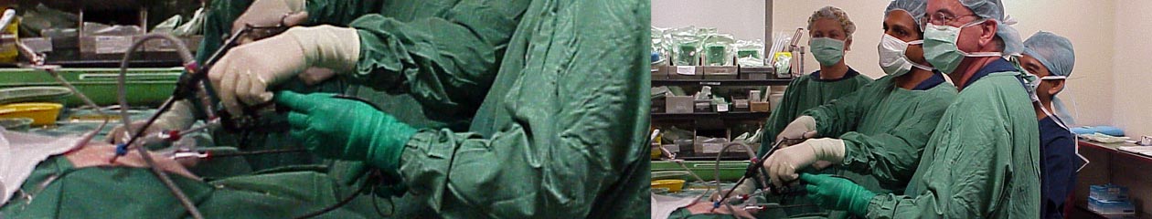
Laparoscopic pyeloplasty offers a minimally invasive treatment option that may be used in patients with either primary or secondary UPJ obstruction and is emerging as a new criterion standard in the treatment of UPJ obstruction [1]. Success rates are comparable with those of open pyeloplasty procedures, and some studies have shown that laparoscopy offers the advantages of decreased morbidity, shorter hospital stay, and quicker recovery. Laparoscopic pyeloplasty is a technically demanding procedure that generally requires significant laparoscopic experience. Robotic-assisted laparoscopic pyeloplasty has become increasingly popular as the robots have become more prevalent. A small intrarenal pelvis is a relative contraindication to laparoscopic pyeloplasty.
References:
- Symons SJ, Bhirud PS, Jain V, Shetty AS, Desai MR. Laparoscopic pyeloplasty: our new gold standard.J Endourol. 2009 Mar. 23(3):463-7.
