Although gallstones have traditionally been considered to be much less common in children than in adults, gallstone disease has increasingly been diagnosed in the pediatric population, mainly owing to the widespread use of ultrasonography [1].
In the past, gallbladder disease in children was usually observed primarily in patients with comorbid conditions such as hemolytic disorders, parenteral nutrition dependence [2], or cystic fibrosis [3] . However, the cholecystectomy rate in children without the diagnosis of hemolytic anemia has doubled in United States in recent years [4] and similar trends are seen in India as well. Obesity is a known risk factor for gallbladder disease, and the increase in the incidence of pediatric gallbladder disease parallels the rise in childhood obesity [5].
If a gallstone obstructs the cystic duct, acute cholecystitis can occur, with distension of the gallbladder wall and possible necrosis and spillage of bile. If gallstones migrate from the gallbladder into the cystic duct and main biliary ductal system, further complications can occur, such as choledocholithiasis, biliary obstruction with or without cholangitis, and gallstone pancreatitis.
The workup of cholelithiasis in pediatric patients is similar to that in adults. The goal is to demonstrate evidence of gall bladder or biliary tract disease. Laboratory tests should include a complete blood count, gamma-glutamyltransferase (GGT), amylase, urinalysis, direct and indirect bilirubin, alkaline phosphatase, and transaminase levels.
All laboratory results in simple cholelithiasis should be within reference ranges. They are of use in identifying more complex disease processes, including biliary obstruction and cholecystitis. Abnormal results on liver function tests or CBC count suggest infection, obstruction, or both.
Ultrasonography of the RUQ is the study of choice in patients with uncomplicated cholelithiasis. Plain radiography, radionuclide scanning, and cholangiopancreatography can also play a role in the assessment of cholelithiasis. Ultrasonography can be used to identify the location of the stone (as demonstrated in the image below, gallbladder wall thickening, the presence of gallbladder sludge, and pericholecystic fluid. Furthermore, an ultrasonographic Murphy sign (expiratory arrest with pressure from the sonographic probe in the RUQ) aids in the diagnosis of cholelithiasis.
No well-established consensus exists regarding the indications for cholecystectomy in asymptomatic pediatric patients [3]. Some authors believe that it is indicated only in patients with comorbidities, particularly hemolytic anemias.
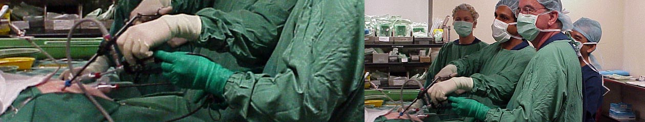
Laparoscopic cholecystectomy is currently the criterion standard in the treatment of symptomatic cholelithiasis. It has been proven to be safe and effective in children, with a low rate of postoperative complications.
In children, when a previous diagnosis of choledocolithiasis or choledochus dilation has been made, ERCP is necessary if obstructive symptoms persist, either before or after an LC [6].
References
- O. Bogue, A.J. Murphy, J.T. Gerstle, et al.Risk factors, complications, and outcomes of gallstones in children: a single-center review.J Pediatr Gastroenterol Nutr, 50 (2010), pp. 303–308
- Wesdorp, D. Bosman, A. de Graaff, et al.Clinical presentations and predisposing factors of cholelithiasis and sludge in children.J Pediatr Gastroenterol Nutr, 31 (2000), pp. 411–417
- Herzog, G. Bouchard. High rate of complicated idiopathic gallstone disease in pediatric patients of a North American tertiary care center. World J Gastroenterol, 14 (2008), pp. 1544–1548
- Balaguer, M. Price, R. Burd. National trends in the utilization of cholecystectomy in children. J Surg Res, 134 (2006), pp. 68–73
- Mehta, M. Lopez, B. Chumpitazi, et al.Clinical characteristics and risk factors for symptomatic pediatric gallbladder disease. Pediatrics, 129 (2012), pp. e82–e88.
- Bonnard A, Seguier-Lipszyc E, Liguory C, et al. Laparoscopic approach as primary treatment of common bile duct stones in children.J Pediatr Surg. 2005 Sep. 40(9):1459-63.
